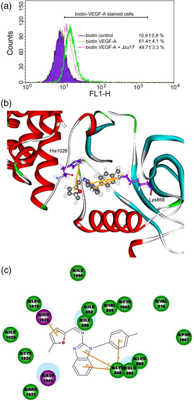
Figure 6.

Molecular docking simulation analysis of Jzu 17. (a)HUVECs were detached, suspended in PBS, and treated with biotin control or biotin VEGF‐A in the absence or presence of Jzu 17. Cells were then treated with avidin‐fluorescein, and the fluorescence derived from biotin VEGF‐A‐stained cells was measured by flow‐cytometry. Results shown are representative of seven independent experiments. (b) Molecular modelling of the interactions between VEGFR‐2 and Jzu 17. The graph shows the docking pose of VEGFR‐2 with Jzu 17. (c) Docking pose of VEGFR‐2 with Jzu 17 using 2D ligand–protein interaction diagrams
