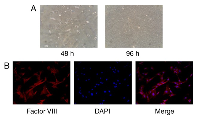
Figure 1.

Morphological and immunological identification of CMECs. Cultures were identified by their morphological features and the expression of factor VIII-related antigen. (A) Primary CMECs possessed a spindle or polygonal shape after 24 h incubation, and a ‘cobblestone’ appearance after 96 h incubation (magnification, ×200). (B) Most of the CMECs were positively stained with a microvascular endothelial cell specific anti-factor VIII antibody (magnification, ×400). CMECs, cardiac microvascular endothelial cells.
