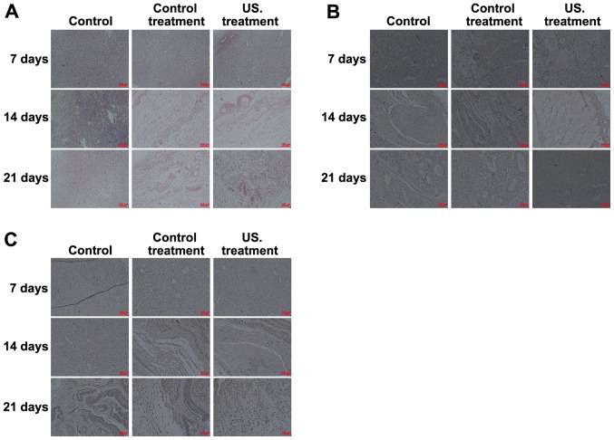
Figure 2.
Histological and IHC analysis of the different groups. (A) Histological staining was performed to investigate the pathological changes occurring in the wound during the healing process. And the red arrow indicates collagen fibers, the blue arrow indicates fibroblasts, and the green arrow indicates neovascularization. IHC analysis indicated that (B) VEGF and (C) TGF-β1 expression significantly increased in the US group compared with the control group. IHC, immunohistochemistry; US, ultrasound.

