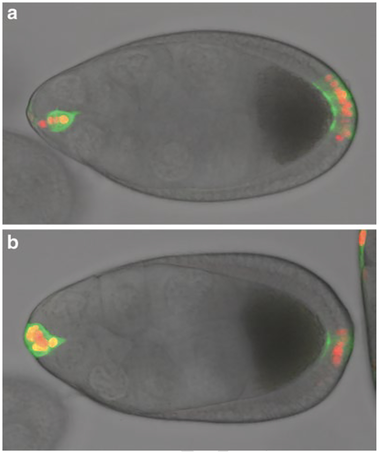
Fig. 5.
DIC images with GFP and nuclear dsRed channels superimposed. (a) Early stage 9 egg chamber with normal morphology and border cell cluster detaching. (b) Stage 9 egg chamber with slightly delayed border cell migration. Note that the outer follicle cells and the proportion of the oocyte indicate that the egg chamber is at a later stage than (a), but the border cells are less detached. In this particular example, the border cells never detached indicating abnormal development

