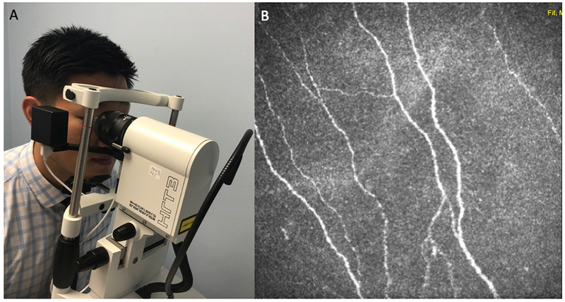
Figure 10:

Imaging Sub-Basal Corneal Nerves with In Vivo Corneal Confocal Microscopy via Heidelberg Retina Tomograph 3 Rostock Cornea Module (Heidelberg, Germany). A: Performing in vivo corneal confocal microscopy. B: Representative output depicting the sub-basal nerve plexus.
