
FIGURE 5.
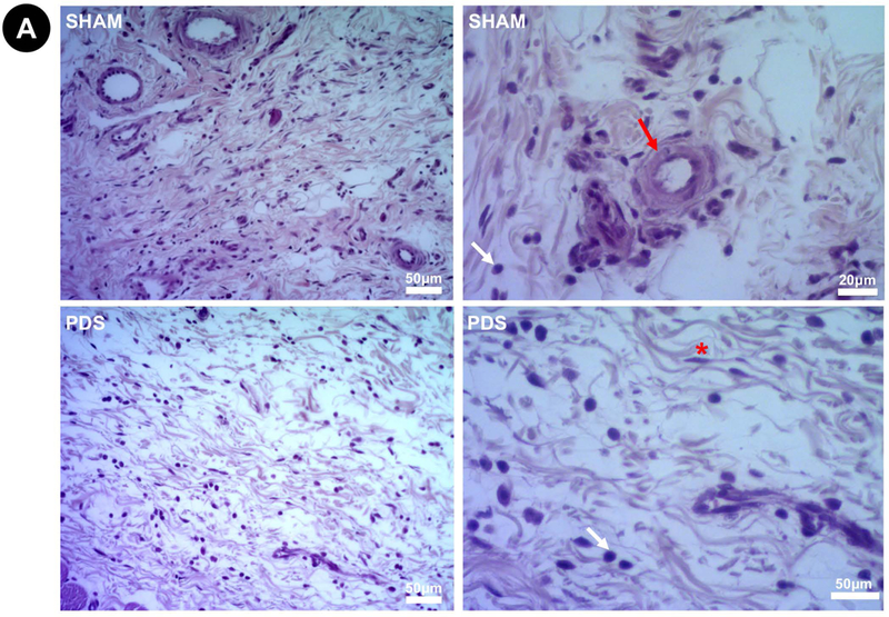
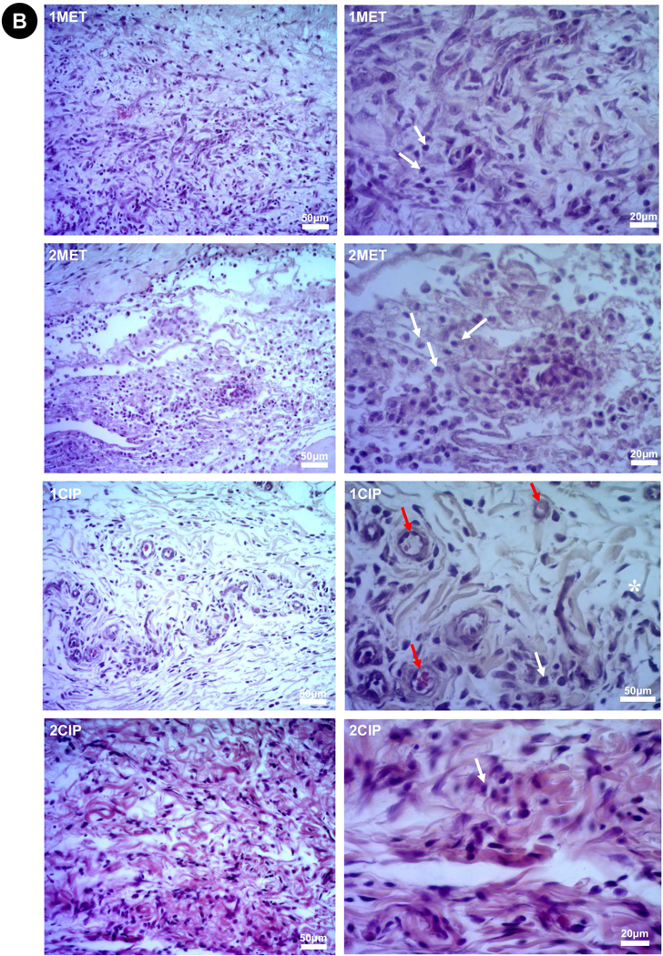
Representative images of H&E stained slides at 3 days (Higher magnifications = 200× left and 400× right). (A) Sham and PDS. (B) 1MET, 1CIP, 2MET, and 2CIP. The degree of inflammatory cell response was classified according to a score system from verbal description using a 4-point scale considering polymorphonuclear leukocytes/mononuclear cells: 0=absent (no inflammatory cells); 1=mild or discrete (few inflammatory cells); 2=moderate (several inflammatory cells and increased reaction zone); and 3=intense or severe (increased reaction zone, occasional foci of neutrophil granulocytes and/or lymphocytes). None of the groups showed severe inflammatory infiltrate. Red arrow (×): blood vessel; white arrow (×): inflammatory cells; asterisk (*): fibroblast.