Fig. 5.
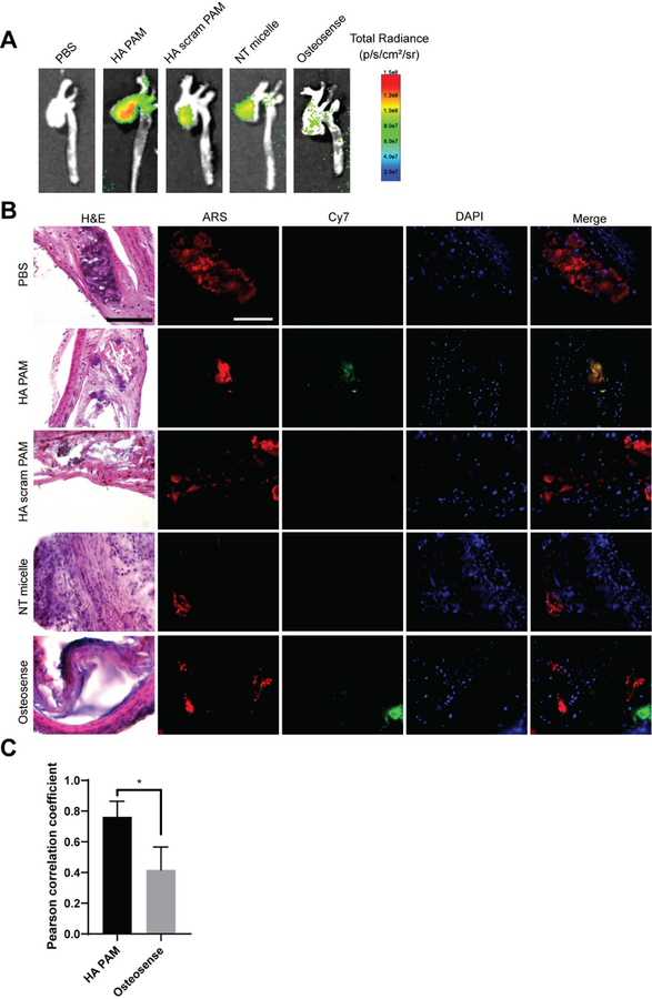
Aortas of ApoE −/− mice 24 hours post-administration of HA PAMs, HA scram PAMs, NT micelles (N=4), Osteosense, or PBS (N=3). A) Ex vivo NIR fluorescence demonstrate enhanced accumulation of HA PAMs compared to other groups. B) Brachiocephalic artery sections stained with H&E show plaque morphology (arrows: calcification). Artery sections were also stained with ARS (red) for calcification and DAPI (blue) for nuclei. Mice treated with HA PAM (green) show colocalization with calcifications (yellow in merged image). No signal is apparent for PBS-, NT micelle-, and HA scram PAM-treated aortas. Osteosense also showed some fluorescence signal in areas of calcification. Scale bar 100 μm. C) Quantification of colocalization of HA PAM or Osteosense with ARS show strong HA PAM colocalization (0.76 ± 0.1) with ARS compared to Osteosense (0.42 ± 0.15). *p≤0.05.

