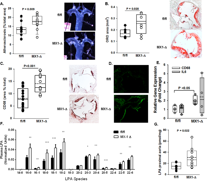
Figure 1. Global reduction in postnatal Plpp3 expression accelerates atherosclerosis.

A) Left Panel: En face analysis of atherosclerosis lesion area (% of arch area) in proximal aortas (n=12–14/group). Right Panel: Representative proximal aortas from fl/fl and MX1-Δ mice on the Ldlr−/− background after 12 weeks on Western diet. B) Oil red-O staining of aortic sinus lesions reported as % area of aortic sinus (n=5–7/group). C) Left Panel: Immunohistological quantification of CD68 from sections of aortic roots from fl/fl and MX1-Δ mice on the Ldlr−/− background after 12 weeks on Western diet (n=12–14/group). Right Panel: Representative images of CD68 staining in the aortic sinus. D) Immunofluorescence staining of ICAM-1 in proximal aortic roots from fl/fl (top) and MX1-Δ (bottom) mice on the Ldlr−/− background after 12 weeks on Western diet. E) Cd68 and Il-6 gene expression (fold change; mean ± SEM) determined by qRT-PCR from proximal aortas of fl/fl and MX1-Δ mice on the Ldlr−/− background after 12 weeks on Western diet (n=4–5/group). F) Plasma LPA species quantification from fl/fl (black bars) and MX1-Δ (white bars) mice on the Ldlr−/− background after 12 weeks on Western diet (n=15/group). *P<0.05, **P<0.01, ***P<0.001. G) Total LPA content (pmol/mg tissue) in proximal aortas from fl/fl and MX1-Δ mice on the Ldlr−/− background 12 weeks after Western diet (n=7–9/group).
