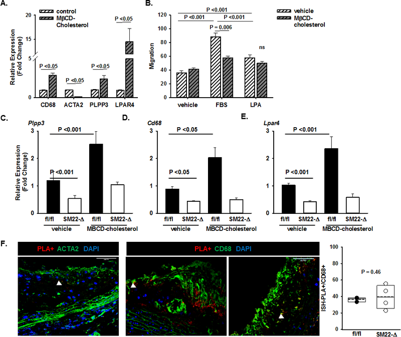
Figure 4. PLPP3 expression is dynamically regulated during SMC phenotypic modulation.

A) Expression of the indicated genes in primary human coronary artery SMC treated with vehicle control or methyl-β-cyclodextrin (MβCD) complexed cholesterol (20μg/mL) for 48 hours to induce modulation towards a foam-cell like phenotype. Relative gene expression (fold change; mean ± SEM) was determined. B) Cell migration to vehicle (DMEM with 0.01% FBS), FBS (DMEM with 10% FBS) or LPA (DMEM with 10μM 18:1 LPA) for 24 hours (right panel; mean numbers of migrated cells ± SEM). Two-way ANOVA was used to determine differences between treatment groups and chemotaxis agent. Relative gene expression (fold change; mean ± SEM) of C) Plpp3, D) Cd68 and E) Lpar4 in primary SMCs from fl/fl and SM22-Δ on the Ldlr−/− background treated with vehicle or MβCD-cholesterol (20μg/mL) for 48 hours. Two-way ANOVA was used to determine differences between treatment groups and genotypes. F) H3K4me2 marker of the MYH11 promoter (red) visualized by ISH-PLA in aortic root sections stained with ACTA (green) and DAPI (blue). Cells positive for the H3K4me2 marker as a percentage of total CD68+ cells (cells/field quantified) in aortic root sections from fl/fl and SM22Δ animals on the Ldlr−/− background fed Western diet for 12 weeks (n=2–4/group).
