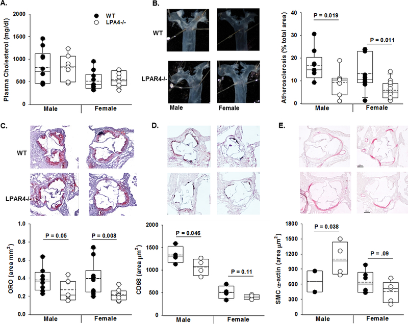
Figure 5. LPAR4 regulates experimental atherosclerosis.

A) Six - 8-week-old wild-type (WT; closed circle) control or Lpar4−/− (open circles) mice were infected with PCSK9 adeno-associated virus. One week later, they were placed on Western diet for 16- weeks and total plasma cholesterol (mg/dL) (n=7–9/group). B) Left Panel: Representative images of proximal aorta. Right Panel: En face analysis of atherosclerosis (% of arch area) in male and female WT (closed circle) and Lpar4−/− (open circles) mice infected with PCSK9 adeno-associated virus and Western diet fed for 16- weeks (n=7–9/group). C) Oil red-O staining and lesion area (% total aorta in root section; n=7–9/group). D) CD68 immunostaining (n=4/group) and E) SM α-actin staining from aortic root sections of male mice (n=2–5/group).
