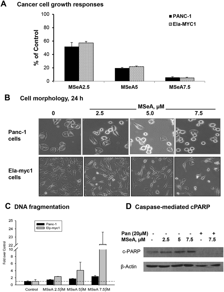
Figure 1. MSeA treatment inhibited cell growth of pancreatic cancer cell lines with variable apoptosis efficacy.
A. Effect of daily MSeA treatment (medium change) on PANC-1 (5 days) and Ela-myc1 (3 days) cell growth. Data were mean ± SEM of triple wells. Absorbance of control wells were set as 100%. B. Representative phase-contrast images of PANC-1 vs. Ela-myc cells after 24 h treatment with MSeA. C. Concentration-dependent induction of apoptotic DNA nucleosomal fragmentation detected by ELISA, after 24 h MSeA exposure, in PANC-1 and Ely-myc1 cell lines. D. Western blot detecting cleaved PARP in PANC-1 cells upon MSeA treatment for 48 h in the presence or absence of pan-caspase inhibitor, zVAD-fmk.

