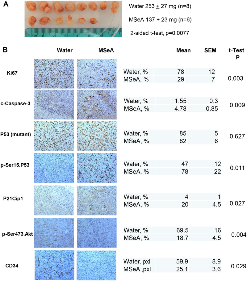
Figure 6. Effect of daily oral-administered MSeA (3 mg Se/kg BW) on growth of subcutaneously inoculated PANC-1 xenografts in SCID mice.
A. The gross appearance of xenograft tumors from SCID mice at termination and the final tumor weights in control and MSeA-treated group. B. Immunohistochemical (IHC) analyses of biomarker changes involved in cell proliferation, apoptosis and angiogenesis in PANC-1 xenograft tumors. Representative images of IHC staining were shown in the left panel and the quantification of the IHC staining was shown in the right panel. Five to 10 pictures were randomly taken under microscope for each section and then analyzed by Image pro plus 6.2 software. For CD34, area in pixels x10−3.

