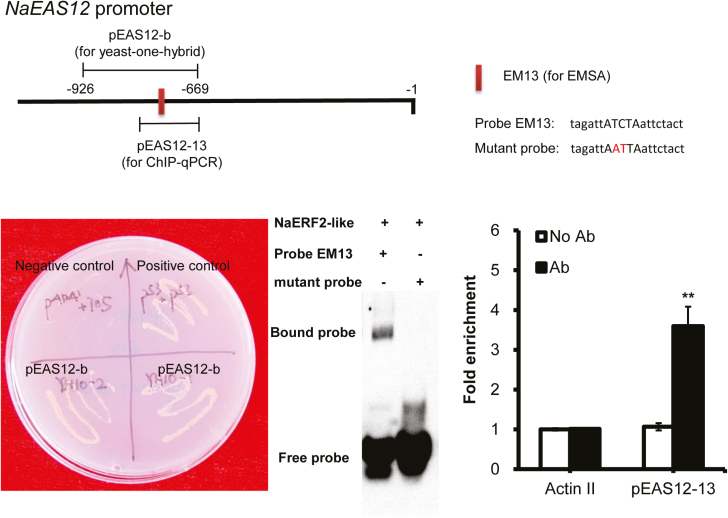
Fig. 9.
The binding of NaERF2-like and NaEAS12 promoter as demonstrated by yeast one-hybrid, EMSA and chromatin immunoprecipitation. The NaEAS12 promoter structure indicated the pEAS12-b (−669 to −926 numbered from the ATG) for yeast one-hybrid, the probe EM13 (5′-tagattATCTaattctact-3′) for EMSA, and the pEAS12-13 (−669 to −781 numbered from the ATG) for ChIP assays. The red letters ‘AT’ indicate the mutated positions. Yeast one-hybrid analysis revealed that NaERF2-like could bind to EAS12-b as the yeast cells could grow on the SD/−His/−Leu medium supplied with 200 ng ml−1 (final concentration) 3-AT; Y1HGold [pGADT7/pEAS12-b-AbAi] was used as a negative control, and Y1HGold [pGADT7 Rec-p53/p53-AbAi] was used as a positive control. EMSA demonstrated that His tagged NaERF2-like could bind to probe EM13 but not to the mutated one. The mutant probe (5′-tagattAATTaattctact-3′) served as a negative control in EMSA. ChIP-real time PCR data indicated NaERF2-like bound to the promoter of NaEAS12. Negative controls were without antibody (no Ab) and with HA antibody but using primers detecting NaActin 2. Asterisks indicate level of significant difference between no Ab and with Ab in pEAS12-13 (Student’s t-test: **P<0.01).

