
Fig 9. Exemplar histology results of sheep brain tissue (‘SH4’).
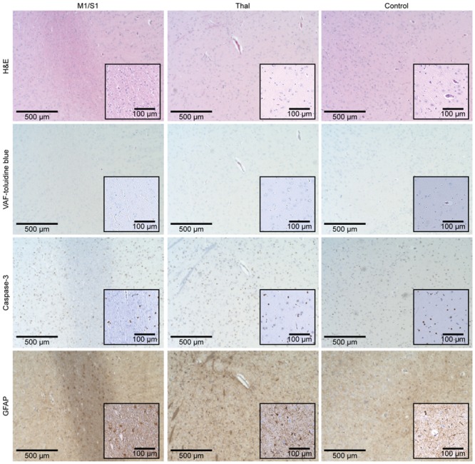
The microscopic images (×40 magnification) of the respective tissue sampling location (M1/S1, Thal, and control site) are displayed according to the staining method (H&E, VAF-toluidine blue, caspase-3, and GFAP). Insets represent magnified images (×100 magnification).