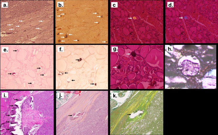
Fig 2. Illustration of the different patterns of crystals deposits in thyroid tissue using optic microscopy.
Crystals located were misdiagnosed by Hematoxylin and Eosin staining (HE) (arrows show the location of colloid crystals that are misdiagnosed by HE staining in (a), but detected by Von Kossa staining as dark composits (arrows show the black staining of crystals with VK coloration inside the vesicles in (b) and also by polarized light microscopy (arrows show the birefringent character of colloid crystals in (c) and (d), magnification x40). Psammoma bodies were detected both by HE (arrows show the HE staining of psammoma bodies outside the vesicles in (e), and Von Kossa staining (arrows show the black staining of psammoma bodies with VK coloration in (f) with no polarized property and (g) psammoma bodies shown with arrows do not have birefringent character). A lamellar aspect was detected on silver coated slide without staining (the arrow show a psammoma body in (h)) (x200 magnification). Large calcification in nodule’s capsule were easily detected with HE (in (i) and (j) capsule calcification are shown with arrows) and also on silver coated slide without staining (an arrow shows in (k) the same capsule calcification than (j) in a silver coated slide without staining) (x40 magnification).

