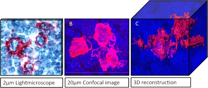
Fig 4. Comparison of imaging techniques in 2D and 3D.
(A) conventional histological CD30 staining in MCcHL. (B) Representation of the same case in three dimensions using a confocal laser scanning microscope. (C) Surface reconstruction of the cell arrangement of a confocal image. The 3D image illustrates the spatial relationship of the cells to each other, their contacts, protrusions and contiguous structures. Images can be rotated in all three axes on the computer.

