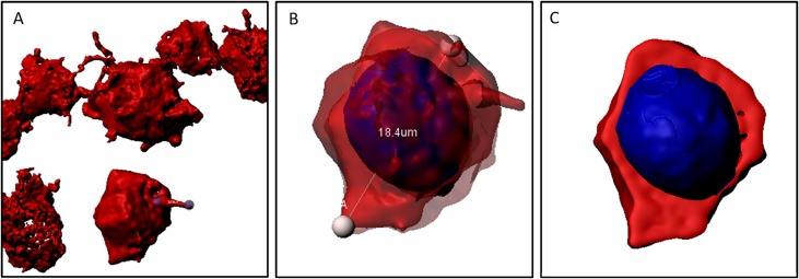
Fig 5.
(A) sample of CD30+ cells of a MCcHL. CD30+ cells are connected by protrusions. (B) shows an excerpt of (A). The cell is semitransparent to determine its diameter (18,4μm) and to shows the nucleus colored in blue. (C) section through the cell. The cytoplasm and the nucleus can be distinguished from each other. The CD30+ proportion (cytoplasm) represents the share we used for calculations.

