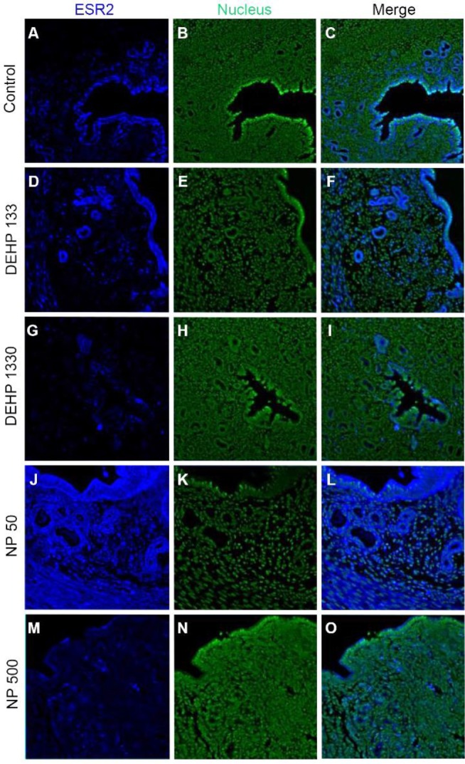
Fig. 3. Tissue specific localization of ESR2 in mouse uterus by DEHP and NP administration. Some of the localized cells came from different tissues of the control. Immunofluorescence was performed with target specific antibodies and the images were analyzed with confocal microscope. (A–C) control, (D–F) 133 μg/L DEHP, (G–I) 1,330 μg/L DEHP, (J–L) 50 μg/L NP, (M–O) 500 μg/L NP. (A, D, G, J, M) ESR2, (B, E, H, K, N) nuclei presented by YOYO-1, (C, F, I, L, O) merged image. ESR2, estrogen receptor 2; DEHP, di(2-ethylhexyl) phthalate; NP, nonylphenol.

