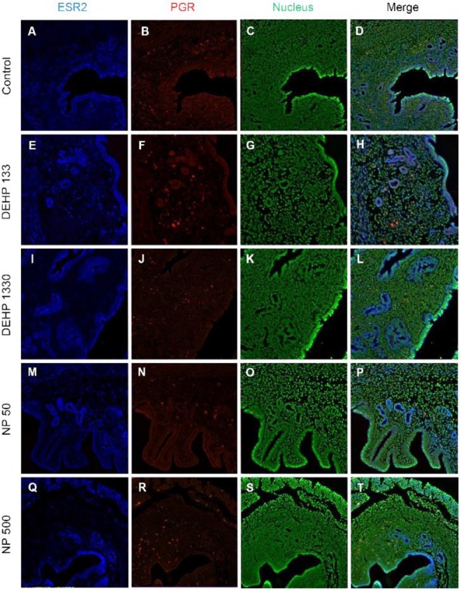
Fig. 7. Microfluorophotography of ESR2 and PGR in mouse uterus. Representative immunofluorescence conducted uteri are shown (magnification, ×100). ESR2 and PGR were not colocalized in the same cells in both stromal and epithelial cells. In 133 μg/L DEHP group, the ESR2 and PGR were colocalized in some epithelial cells. (A–D) control, (E–H) DEHP 133 μg/L, (I–L) DEHP 1,330 μg/L, (M–P) NP 50 μg/L, (Q–T) NP 500 μg/L. (A, E, I, M, Q) ESR2, (B, F, J, N, R) PGR, (C, G, K, O, S) nuclei presented by YOYO-1, (D, H, L, P, T) merged photomicrograph. ESR2, estrogen receptor 2; PGR, progesterone receptor; DEHP, di(2-ethylhexyl) phthalate; NP, nonylphenol.