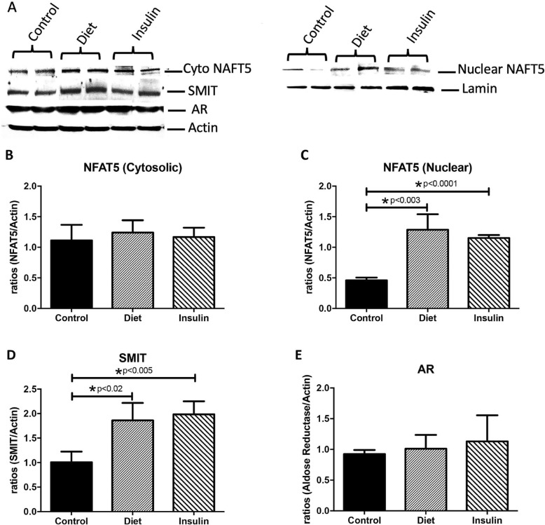
Fig. 3.
NFAT5, SMIT and AR in control and GDM human placentas. Cytosolic and nuclear levels of NFAT5, SMIT and AR (n = 5) were measured by western blot and quantified by Spot Denso analysis. Characteristic western blots for NFAT5, SMIT and AR are shown in (a). Cytosolic NAFT5 levels were not changed in the GDM-D or the GDM-I placentas when compared to control samples (b). Nuclear NAFT5 levels were elevated in in both GDM-D and GDM-I (p < 0.05) placenta when compared to control placenta samples (c). Cytosolic SMIT was increased in both GDM-D and GDM-I placenta as compared to controls (d). There were no changes for AR expression between control and GDM placentas (e). Experiments were conducted in triplicate and statistically different values are noted as * p < 0.05

