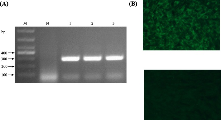
Fig. 3.
Characterization of TMUV isolates. a Detection of TMUV Y by RT-PCR. Lane M, molecular weight marker; lane N, negative control; lanes 1–3 indicate the third to fifth passages of Y virus respectively. b Identification of TMUV GL by IFA. Top, BHK-21 cells infected with TMUV GL; bottom, negative control

