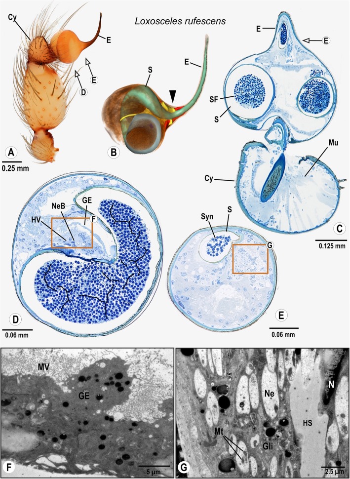
Fig. 5.
Palpal organ of Loxosceles rufescens; external morphology (a), histology (c–e) ultrastructure as documented by TEM (f, g), and 3D reconstruction of the spermophor (green), nervous tissue (yellow) and distinct cell clusters/“sensor epidermal tissue” (red) as based on segmentation of histological image stacks (b). Arrows indicate planes chosen for semithin sections (a) and the arrowhead marks the terminals of the bulb nerve (b). Box in (d) indicates the location of glandular tissue in the bulb, ultrastructural details are given in (F). Box in (e) marks the branches of the bulb nerve, highly magnified in G. Abbreviations: Cy Cymbium, E Embolus, GE Glandular epithelium, Gli Glial cell processes, HS Haemolymph space HV Haemolymph Vessel, Mu Muscle, Mt Mitochondria, MV Brush of microvilli, Ne Neurite, NeB Neurite Bundle, N Nucleus, S Spermophor, SF Seminal fluid, Syn Synspermium

