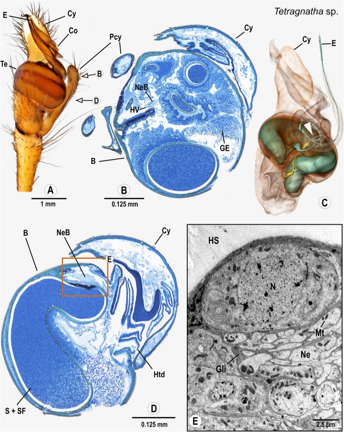
Fig. 8.
Palpal organ of Tetragnatha montana.; external morphology showing the general organization (a) as well as histology (b, d), ultrastructure as documented by TEM (e), and 3D reconstruction of the spermophor (green) and nervous tissue (yellow) of Tetragnatha extensa as based on segmentation of histological image stacks (c). Arrows in (a) indicate planes for semi-thin cross sections taken at distal end (b) and midlevel (e) of the bulb. Arrowhead marks terminals of bulb nerve (c). Note that the embolus in (a) rests in a ridge of the conductor and therefore differs from the one depicted in (c). Box shows a sector where neuronal somata and a neurite bundle branched off the bulb nerve are present, part of this sector is shown in (e) magnified to ultrastructural level. Abbreviations: B Bulbus, Co Conductor, Cy Cymbium, E Embolus, GE Glandular epithelium, Gli Glial cell processes, HS Haemolymph space, Htd Haematodocha, HV Haemolymph Vessel, Mt Mitochondria, N Nucleus, Ne Neurite, NeB Neurite Bundle, Pcy Paracymbium, S Spermophor, SF Seminal fluid, Te Tegulum

