. 2019 Oct 17;60(11):1005–1012. doi: 10.3349/ymj.2019.60.11.1005

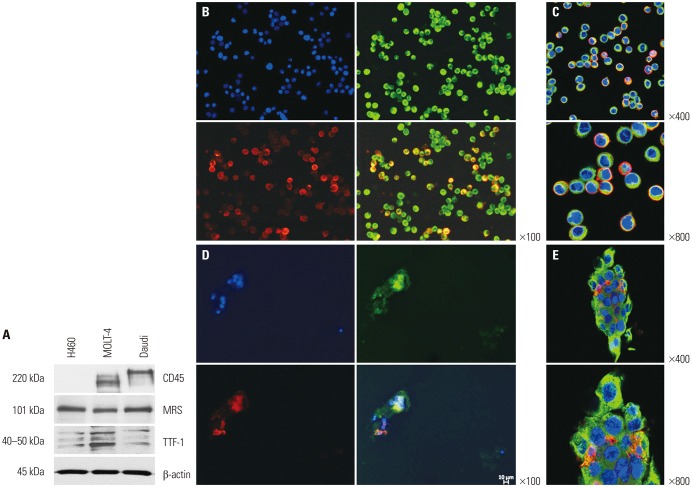
Fig. 3. Immunoblotting of methionyl-tRNA synthetase (MRS)-positive H460 cells, CD45/CD3-positive Molt-4 cells, and CD45/CD20-positive Daudi cells (A). Fluorescent immunochemical staining for MRS and CD45 in the reference samples containing a fixed ratio of H460, Daudi, and Molt-4 cells (B and C) and endobronchial ultrasound-guided transbronchial needle aspirate-derived samples (D and E). Images were taken using a fluorescent microscope (B and D) and confocal microscope (C and E). Blue represents nucleus (4′, 6-Diamidine-2′-phenylindole dihydrochloride: DAPI); green, MRS; and red, CD45. B and D confocal images show merged images only. TTF-1, thyroid transcription factor 1.

Fig. 3