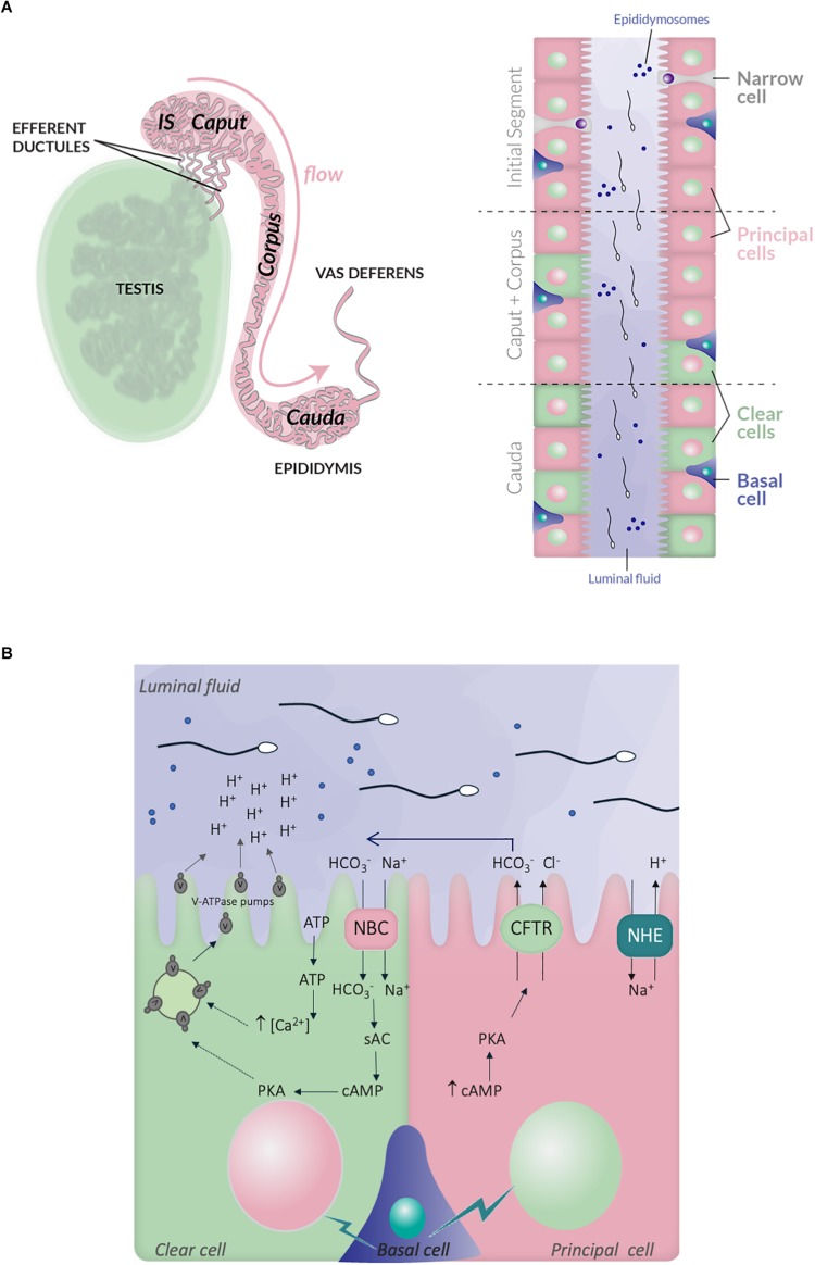
FIGURE 2.
Schematic representation of the epididymis structure and ionic exchanges between epithelial cells, which control luminal acidification. (A) Left panel, the testis, efferent ductules and epididymis are schematized. The different regions within the epididymis, in mouse, are indicated: initial segment, caput, corpus and cauda, following the proximal-distal axis. Right panel, the distribution of the different epithelial cell-types within the epididymal tract is also illustrated: principal cells (PCs), clear cells (CCs), narrow cells (NCs), and basal cells; the luminal fluid shows epididymosomes, which are small vesicles transferring material from epithelia cells to the sperm cells. (B) Simplified representation of the main ionic fluxes and cross talks occuring between principal, clear, and basal cells. CCs expressed the V-ATPase pumps, which expression at the plasma membrane is induced by HCO3– and c-AMP dependent pathway. The HCO3– influx in CCs is mediated by the NBC sodium- HCO3– transporter. ATP also induces intracellular rise of Ca2+, which increase V-ATPase translocation at the plasma membrane and proton secretion. PCs express the NHE3 sodium-proton antiporter, which contributes to proton secretion and luminal acidification. They also secrete HCO3– through the CFTR channel. Lastly, basal cells transmit physiological cues, in particular during sexual arousal, which regulate the activity of principal and CCs.

