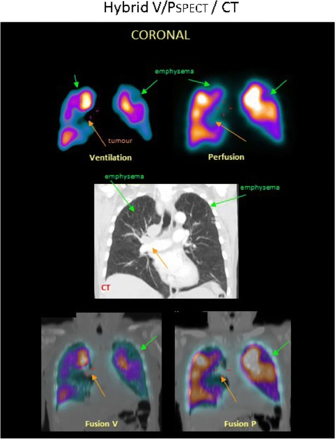
Fig. 6.

A patient with COPD, emphysema and tumour. Coronal slices display uneven distribution of ventilation with a pattern of deposition of 99mTc-Technegas® that is typical for COPD. Perfusion follows the ventilation pattern. Matched ventilation and perfusion defects are observed in both upper lobes (green arrows) and to the right of the mediastinum (orange arrows). In the medial row of the corresponding coronal CT slice, emphysema is seen in both upper lobes (green arrows), as is a tumour in the mediastinum (orange arrow). Fusion images of CT and ventilation SPECT and CT and perfusion SPECT are shown
