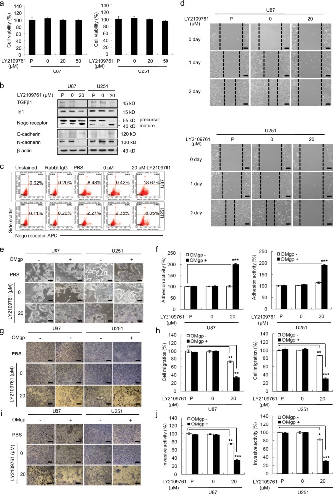
Fig. 2. Role of mature NgR on cellular behavior in LY2109761-treated GBM.
a WST-1 assay revealed that the cell viability was not affected by LY2109761 treatment in U87 and U251 cells. b Western blot analysis showing the expression levels of TGFβ1, Nogo receptor, and E-cadherin in LY2109761-treated U87 and U251 cells. The expression levels of all proteins were normalized to those of β-actin. c FACS analysis showing that the inhibition of TGFβ1 resulted in increases in mature surface Nogo receptor. d The scratch–wound migration activity of LY2109761-treated U87 and U251 cells, as determined by the scratch–wound migration assay. U87 and U251 cells were treated with 20 μM LY2109761 for 48 h prior to the assay, and the wound was created by scratching using a sterile 200-μl pipette tip. Duplicate wells were used for each condition, and three fields per well were captured at each time point over a period of 48 h. Images of the same fields were taken at days 0–2 (×100 magnification). Black scale bar = 100 μm. The inhibition of TGFβ1 suppressed the migratory ability of U87 and U251 cells. e The adhesion activity of LY2109761-treated U87 and U251 cells, as determined by an OMgp-coated matrix adhesion assay. Phase-contrast image of U87 and U251 cells treated with LY2109761, showing representative cells adhering to the well with or without OMgp (100 ng/ml) (×100 magnification). Black scale bar = 100 μm. f Quantification of adhesive U87 and U251 cells treated with LY2109761. The inhibition of TGFβ1 increased the adhesion activity of U87 and U251 cells through the upregulation of OMgp responsiveness. g Cell migration activity of LY2109761-treated U87 and U251 cells, as determined by an OMgp-treated transwell migration assay. U87 and U251 cells were treated with LY2109761 for 48 h before the assay. The upper chamber of the transwells was seeded with 0.2 ml of cells (4 × 10⁵ cells/ml) in medium with 5% FBS supplemented with half the amount of the treatment only or with OMgp (100 ng/ml), and 0.6 ml of DMEM containing 20% FBS was added to the lower chambers. After 24 h, migrating cells were stained with crystal violet, and images were taken (×100 magnification). Black scale bar = 100 μm. h The migrating cells were counted under a microscope in three different fields per experiment. i Cell invasion was examined through a membrane filter coated with OMgp/Matrigel or Matrigel alone. After 24 h, invading cells were stained with crystal violet, and images were taken (×100 magnification). Black scale bar = 100 μm. j The invading cells were counted under a microscope in three different fields per experiment. The mean values and the standard error were obtained from three individual experiments. *p < 0.01, **p < 0.005, and ***p < 0.001

