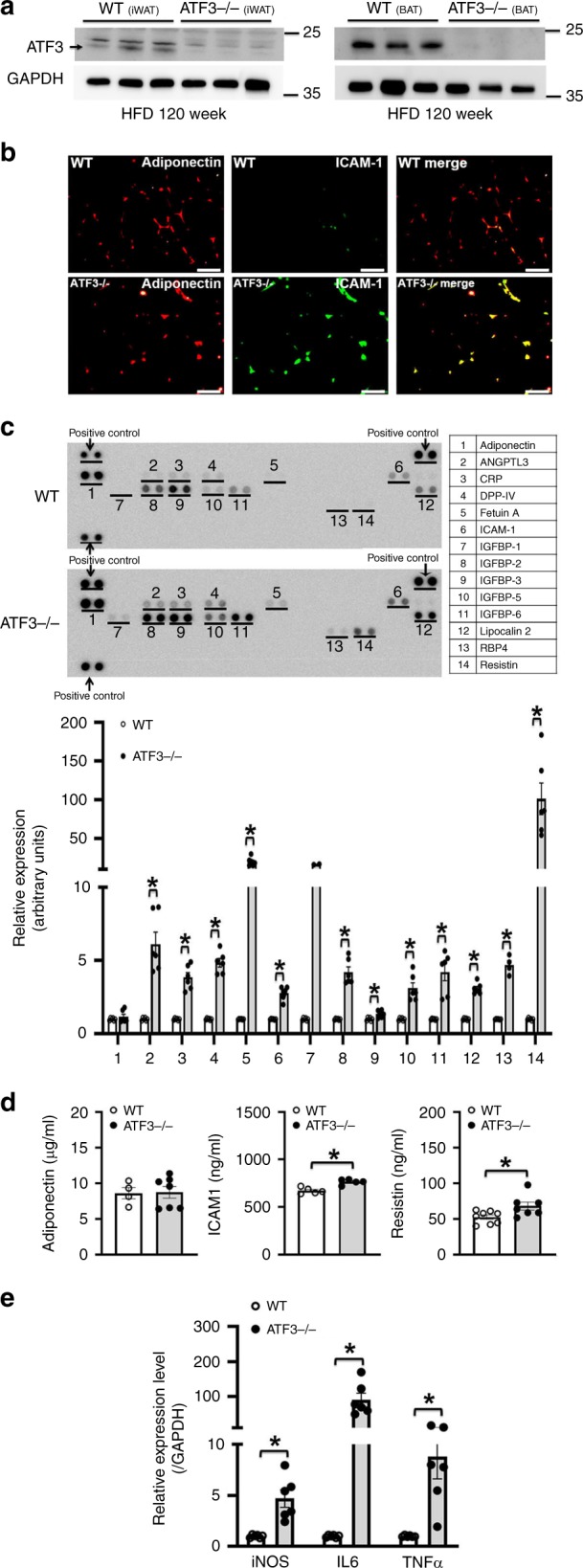
Fig. 3.

Loss of ATF3 aggravated the expression of inflammation-related genes in HFD-induced obese mice. a ATF3 protein level in iWAT and BAT of wild-type and ATF3−/− mice after HFD feeding for 12 weeks. b Representative immunofluorescence images of adiponectin (red IF) and ICAM-1 (green IF) in wild-type and ATF3−/− mice. Yellow scale bar indicated the size of adipocyte tissues. c Serum protein levels of adipokine and inflammation-related genes in wild-type and ATF3−/− mice after HFD feeding for 8 weeks by adipokine assays; Gel-Pro Analyzer software was used for densitometry of blots. d Serum protein level of adiponectin, ICAM-1 and resistin by ELISA assays in wild-type and ATF3−/−mice after HFD feeding for 8 weeks. e Quantified real-time PCR analysis of mRNA levels of iNOS, IL-6, and TNFα in livers of wild-type and ATF3−/− mice. For a–c, n = 3 per group. For d, wild-type (n = 4 in adiponectin; n = 5 in ICAM1; n = 8 in resistin), ATF3−/− (n = 7 in adiponectin; n = 5 in ICAM1; n = 7 in resistin). For e, n = 6 per group. Scale bar for image b: 50 µm. Data are mean ± SEM; *p < 0.05 compared to wild-type
