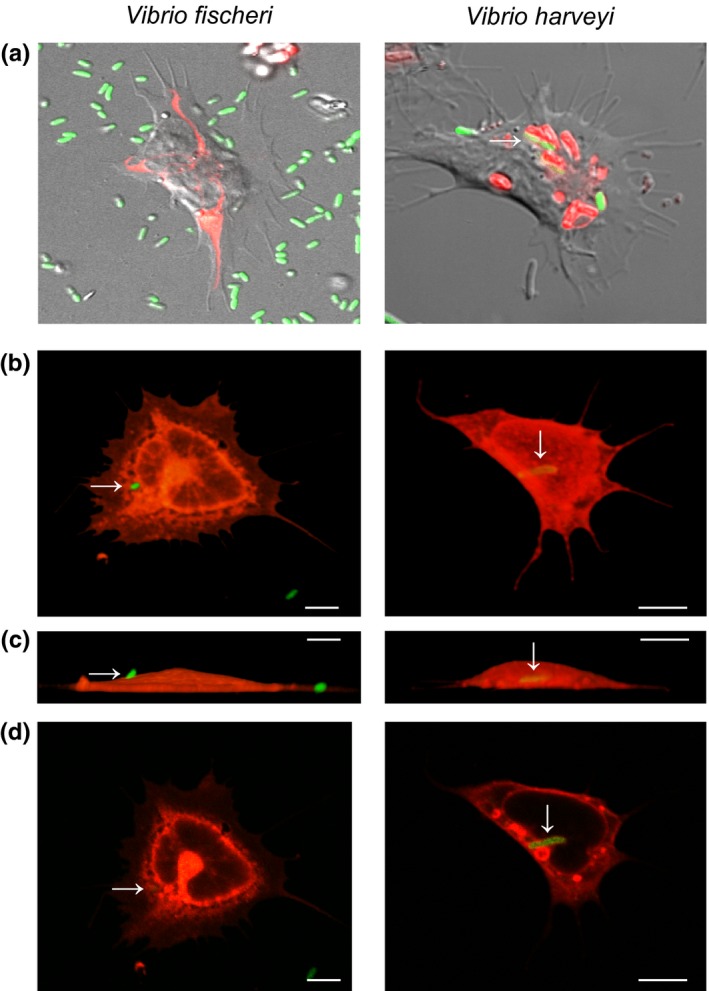
Figure 3.

Phagocytosis of Vibrio fischeri and V. harveyi by juvenile hemocytes. (a) DIC confocal images of live juvenile hemocytes bound to and/or internalizing GFP‐expressing bacteria (green), and colocalized with acidic compartments (red, pHrodo pH indicator). (b–d) Confocal images of fixed juvenile hemocytes (red, Con A) bound to and/or internalizing GFP‐expressing bacteria (arrows, green), scale, 10 µm. (b) Top view, 3D images. (c) Side view, 3D images, with the adherent side of the hemocyte at the bottom of the images. (d) Image section through the middle of the cell, to confirm internalization of bacteria
