
Figure 5.
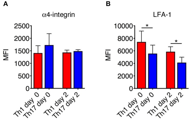
Integrin expression on Th1 and Th17 cells. The expression of α4 integrins (A) and LFA-1 (B) on Th1 and Th17 was evaluated both in vitro before injection and ex vivo on exogenous CMAC+ T cells isolated from spinal cords 48 h after adoptive transfer. Samples were analyzed by flow cytometry. The mean fluorescence intensity (MIF) of LFA-1 was statistically higher on Th1 cells than Th17 cells both before injection and 48 h after cell transfer. Data represent the mean ± SD of four mice per condition from two independent experiments (*P < 0.05).