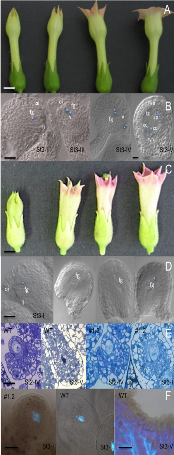
Figure 6.

Detailed type I MYB10.1 phenotype during flower development. (A) Alteration of floral development in type I compared to (C) WT (stages were defined according to Koltunow et al. (1990). Bars = 200 pixel. (B) Cleared sections in wild-type developing ovules from stage st3-I to st3-V, the seven-celled embryo sac. (E) In ovules of overexpressing lines, the development is blocked to st3-I. (D) Thin sections of WT and type I ovules confirm the developmental arrest. (F) Aniline blue staining evidenced the persisting deposition of callose in pollinated type I plants, while the callose disappears in pollinated wild-type flowers. Ovules stages are according to Schneitz et al. (1995). ap, antipodal cells; cc, central cell; ec, egg cell; fg, FG; ii, inner integument; oi, outer integument; syn, synergid cells; v, vacuole. Bars are 1 cm in A and B; 50 µm in B, D, E, F St3-I; 200 µm in F St3-V.
