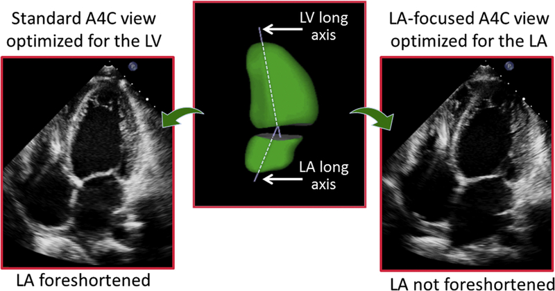
Fig. 5.
Example of an apical 4-chamber view, optimized to depict maximal length of the LV (left). In this view, the LA is foreshortened, in contrast with an LA-focused view specifically optimized to visualize the atrium at its maximal length (right). Atrial foreshortening occurs because the long axes of the ventricle are not the same, as depicted in this 3D reconstruction of both left heart chambers (center).

