Figure 5.
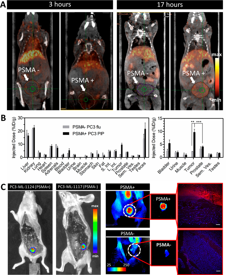
64Cu-LC-Pyro-enabled PET imaging in an orthotopic prostate cancer model and fluorescence detection of PSMA+ micrometastases with LC-Pyro. (A) Sagittal PET/CT images from one mouse bearing either a PSMA+ PC3 PIP or PSMA- PC3 flu orthotopic prostate tumor at 3 and 17 h after intravenous administration of 64Cu -LC-Pyro. (B) 64Cu-labeled LC-Pyro biodistribution in the tumors and the surrounding organs quantified via gamma counting (n = 4 for PSMA+ PC3 PIP; n = 3 for PSMA- PC3 flu, **P < 0.01; ***P < 0.001). (C) In situ bioluminescence images of mice bearing PSMA+ (PC3-ML-1124) and PSMA- (PC3-ML-1117) metastatic nodules (internal organs removed, to expose retroperitoneal cavity). Corresponding fluorescence images demonstrating specific uptake of LC-Pyro in the PSMA+ nodule and the fluorescence microscopic analysis of 10 μm frozen sections (LC-Pyro, red; DAPI, blue; scale = 20 μm).

