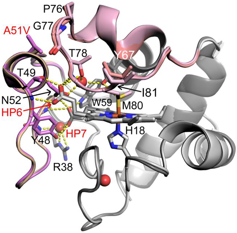
Figure 1.
Overlay of the A51V (PDB ID: 6DUJ, chain A, dark gray) and WT (PBD ID: 3ZCF, chain A, light gray) Hu Cytc structures. Ω-loop C (residues 40 – 57) is shown in wheat (WT) and light purple (A51V). Ω-loop D (residues 70 – 85) is shown in light pink (A51V) and salmon (WT). Stick models are used for the heme and selected side chains. Buried water molecules are shown as red spheres. Hydrogen bonds are shown as yellow dashed lines.

