
Fig. 1: MBV isolated from ECM bioscaffolds contain full-length (32 kDa) IL-33.
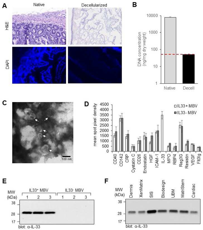
(A, B) Decellularization efficacy. The presence of nuclei in native mouse small intestine tissue and decellularized mouse intestine tissue was assessed by hemotoxylin and eosin (H&E) staining and 4’,6-diamidino-2-phenylindole (DAPI) staining (A). Concentration of double stranded DNA was quantified using the PicoGreen assay; y-axis is log scale; n = 4; p <0.005. Red line denotes established DNA criteria against which decellularization efficacy was verified. Native dsDNA concentration = 7747.34 ± 748; Decellularized dsDNA concentration = 49.87 ± 4.84 (B). (C) Transmission electron microscopy imaging of MBV isolated from decellularized mouse intestine. (D) Graphic representation of the quantitation of 15 cytokines with the highest expression levels in MBV isolated from decellularized wt and il33−/− mouse intestines. Shown are mean ± s.d, n=3 biological replicates. (E) Immunoblot analysis of IL-33 expression levels in MBV isolated from three decellularized wt mouse intestines (IL33+ MBV) or three decellularized il33−/− mouse intestines (IL33- MBV). (F) Immunoblot analysis of IL-33 expression levels in MBV isolated from laboratory produced porcine urinary bladder matrix (UBM), small intestinal submucosa (SIS), dermis, and cardiac ECM; and three commercially available ECM bioscaffold equivalents: ACell® MatriStem™ (porcine UBM), BD® XenMatrix™ (porcine dermis), and Cook Biotech® Biodesign™ (porcine SIS).