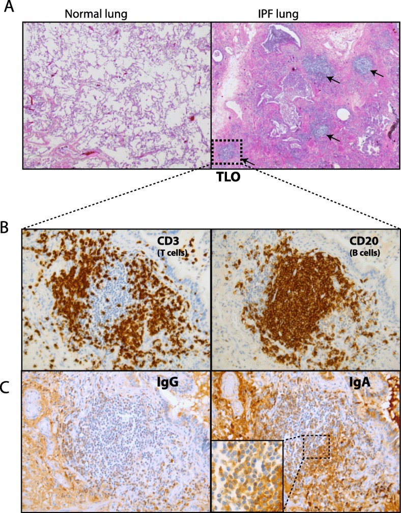
Fig. 3.

IgA+ B cells are present within TLO structures of IPF lungs. (a) Hematoxylin and eosin (h&e) staining of a control lung and IPF lung showing numerous TLOs (black arrows). (b) Pulmonary TLOs of IPF lung stained with anti-CD3 (T cells) and anti-CD20 (B-cells). (c) Representative images of staining with anti-IgG and anti-IgA. Magnification: 10x (a) and 40x (b and c)
