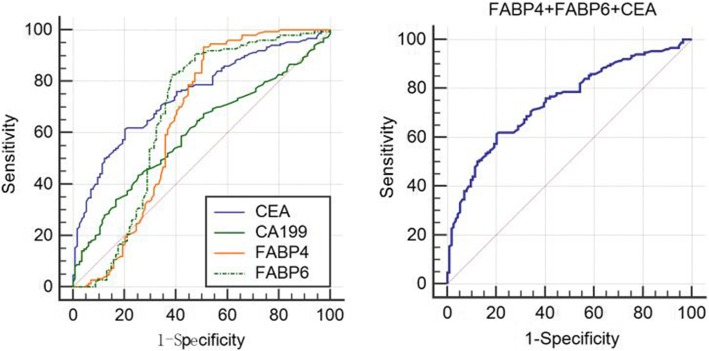
Fig. 4.
Receiver operating characteristics curve analysis using serum FABP4, FABP6,CEA, and CA199 in CRC, respectively (Left). Joint detection of FABP4, FABP, and CEA in CRC for discriminating CRC from normal subjects (Right). ROC curve analyses showed that the ROC curve areas for FABP4, FABP6, and CEA as well CA19-9 in CRC are 0.658 (95%CI 0.598–0.714), 0.683 (95%CI 0.624–0.738), 0.689 (95%CI 0.631–0.744), 0.592 (95%CI 0.531–0.651), respectively. The optimal sensitivity and specificity obtained by movement of the cutoff value of serum FABP4, which was 223.35 pg/ml, were 93.20% (95%CI 87.8–96.7) and 48.8% (95%CI 39.8–57.9) in discriminating CRC from the normal control. Similarly, the optimal sensitivity and specificity obtained by movement of the cutoff value of serum FABP6, which was 347.26 pg/ml, were 83.70% (95%CI 76.7–89.3) and 58.4% (95%CI 49.2–67.1) in discriminating CRC from the normal control. The optimal sensitivity and specificity obtained by movement of the cutoff value of serum CEA, which was 7.5 ng/ml, were 53.06% (95%CI 44.7–61.3) and 77.60% (95%CI 69.3–84.6) in discriminating CRC from the normal control, and the optimal sensitivity and specificity obtained by movement of the cutoff value of serum CA19-9, which was 14.24 U/ml, were 46.26% (95%CI 38.0–54.7) and 68.80% (95%CI 59.9–76.8) in discriminating CRC from the normal control. When combined detection of FABP4, FABP6, and CEA, the area of ROC curves is 0.746 (95% CI 0.689–0.798), and the optimal sensitivity and specificity were 61.33% (53.0–69.2) and 79.82% (71.3–86.8), respectively. Diagonal segments are produced by ties

