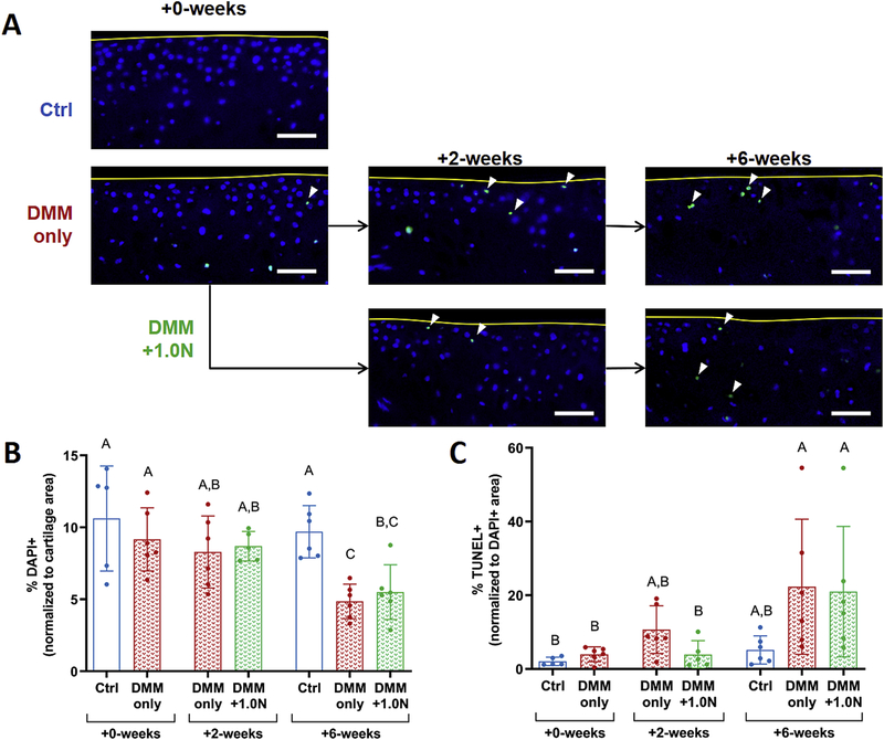
Figure 5.
Low-level cyclic compression had subtle beneficial effects on cellularity and apoptosis. (A) Representative DAPI (blue) overlaid with TUNEL (green) images show chondrocyte loss and apoptosis following DMM surgery, with the greatest degree of cell loss in the +6-week DMM-only group. (B) Cellularity decreased in the +6-week DMM-only group compared to the +2-week DMM-only limbs, whereas cellularity in the +6-week DMM+1.0N group was not different from the +2-week timepoint. However, cell loss was not different between the and DMM-only and DMM+1.0Ngroups at the +2-week and +6-week time points. (C) Chondrocyte apoptosis increased after DMM surgery. At +2-weeks, apoptosis levels in the DMM-only group were not different from +6-week levels, whereas the increase in apoptosis in the DMM+1.0N group was delayed. However, chondrocyte apoptosis was not different between the DMM-only and DMM+1.0N groups at the +2-week and +6-week time points. Scale bars = 100 pm. Arrow heads denote apoptotic cells. Yellow curves designate tibial cartilage surface. Mean ± SD shown with individual data points overlaid. Different letters between bars indicate significant differences in the means by one-factor ANOVA, followed by Tukey post-hoc tests (p<0.05).

