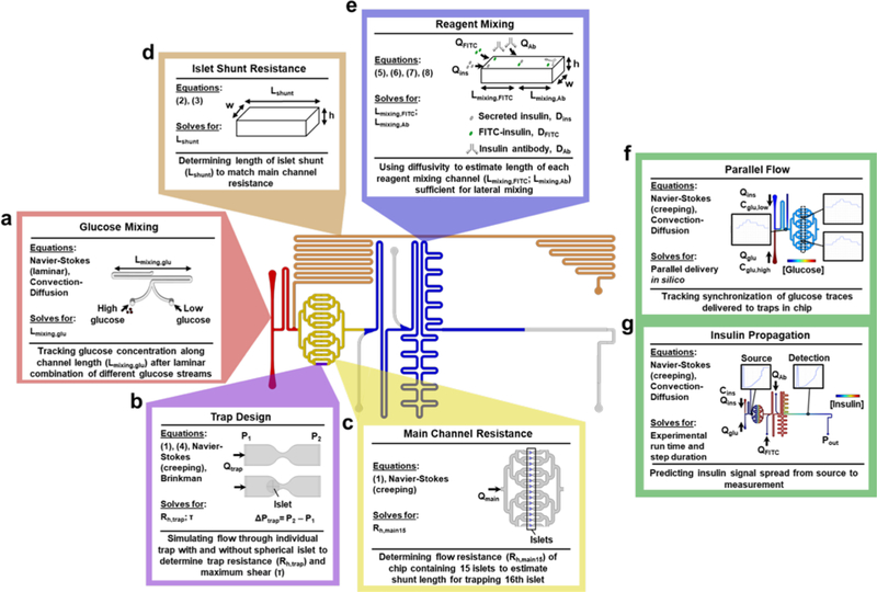
Figure 2 |. Modeling for chip feature design.
Graphical schematic outlining the model design for the ‘Islet on a Chip’ with specific regions highlighted. a. Glucose mixing channel. Three-dimensional flow and convection-diffusion modeled using CAD to ensure sufficient channel length (Lmixing,glu) for complete mixing of glucose input streams. b. Islet trap. Single traps were modeled with three-dimensional flow simulations to predict changes in pressure (ΔP), flow rate (Qtrap), resistance, and shear (τ) in the presence and absence of islets. Traps were designed to maximize inward flow, while retaining minimal shear. c. Main channels. Hydraulic resistance (Rmain) through main chip channels was modeled with 15 occupied traps in two-dimensions using combined glucose and islet inlet flow (Qmain). d. Islet shunt. A rectangular channel with height (h) and width (w) was resistance-matched to Rmain by setting its length (Lshunt) e. Reagent mixing channels. The lengths of the reagent mixing channels (Lmixing ,FITC and Lmixing ,Ab) were determined using the molecular diffusivities of fluorescent reagents (DFITC and DAb) and inlet flow rates (Qins, QFITC, and QAb). f. Parallel flow. For total chip design, delivery of glucose pulses on the chip were simulated with a time-dependent, two-dimensional model of flow and convection-diffusion. Low (cglu,low) and high (cglu,high) concentrations of glucose were delivered at variable flow rates from the glucose and islet inlets (Qglu, Qins). Graphic depicts the spatial concentration map of glucose at one time point of simulation of insulin and insets show traces of glucose concentrations at different locations on the chip. g. Insulin propagation. Model from panel f adapted to simulate insulin propagation in the chip based on a fixed concentration of insulin delivered from the islet inlet (cins). Flow from the regent inlets was also simulated (QFITC, QAb), with outlet pressure (Pout) matched to atmospheric pressure. Graphic depicts the spatial concentration map of insulin at one time point of simulation of insulin and insets show concentration traces at the source and detection point.

