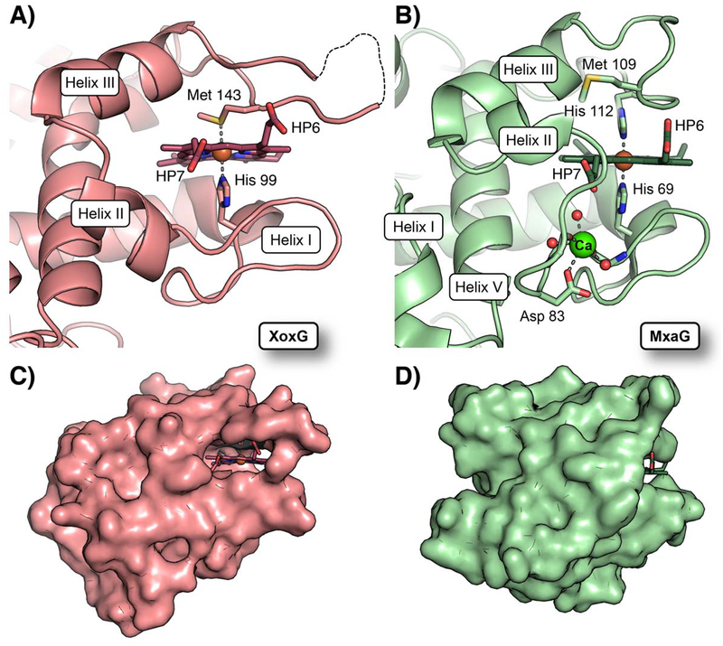
Figure 5. Comparison of heme solvent exposure in XoxG and MxaG.
(A) A ribbon diagram of the XoxG structure near the heme cofactor. The heme is highly solvent exposed because helix II is more extended and it resides further away from the heme propionate face. Heme propionate substituents are designated HP6 and HP7. (B) A ribbon diagram of MxaG (PDB ID: 2C8S) in the vicinity of the heme reveals that Helix II unwinds to coordinate a CaII (green sphere) via the side chain of Asp 83, backbone carbonyls of Tyr 85 and Gly 80, and three water molecules (red spheres). This structural change positions a loop in front of the inner propionate ligand. An FeIII ion (orange sphere) is bound to His 69 and His 112 in the crystal structure, but the latter ligand is replaced by Met 109 in solution. A surface representation of XoxG (C) and MxaG (D) shows that both heme propionate (HP) groups are exposed in XoxG, whereas the inner propionate of MxaG is buried. Increased access to solvent in XoxG should render the heme more easily reduced.

