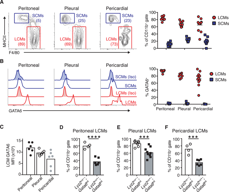
Figure 1.
Resident cavity macrophages express and depend on GATA6. (A) Representative gating and quantification of large cavity macrophages (LCMs) and small cavity macrophages (SCMs) and in peritoneal, pleural and pericardial cavities. Numbers are frequency of cells in gate. (B) Intracellular staining and quantification of GATA6 levels in LCMs and SCMs in peritoneal, pleural and pericardial cavities. Unshaded histograms represent isotype staining and shaded histograms are GATA6 staining. (C) LCM GATA6 mean fluorescence intensity (MFI) in peritoneal, pleural and pericardial cavities. (D-F) Frequency of LCMs in peritoneal (C), pleural (D) and pericardial cavities (E) in Lyz2cre-;Gata6fl/fl or Lyz2cre+;Gata6fl/fl mice. (A-C) Cells were gated as CD45+CD19-Gr1-SiglecF- CD11b+. (D-F) Cells were gated as CD11b+ICAM2+. Data are representative or from of ≥3 (A-E) or 1 (F) experiments. Each dot represents 1 mouse. Mean values are shown. *p<0.05, **p<0.005, ***p<0.0005, ****p<0.0001, as determined by unpaired students T-test.

