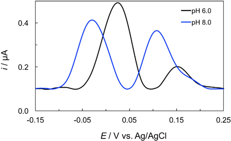
Figure 5.
DPVs of 25.0 μM NE and 10.0 μM 5-HT in 0.1 M PB on Janus-ePAD at WE1 and WE2 where pH of 0.1 M PBS was simultaneously in situ adjusted from pH 7.0 to pH 6.0 (WE1) and 8.0 (WE2), respectively. DPV measurements were performed at an amplitude of 60 mV, potential increment of 4 Hz, and a pulse width of 0.05 s.

