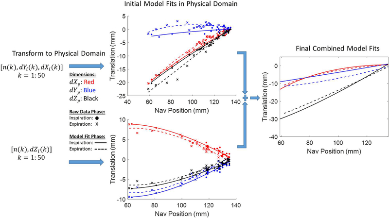
Figure 3.
Motion extraction from training data produces sets of [n(k), dYi(k), dXi(k)] from the SAX view and [n(k), dZi(k)] from the 2CH view for k = 1:50, where n(k) represents the navigator position and dXi(k), dYi(k), dZi(k) the position of the heart in the image coordinate system. Each set is classified as inspiratory or expiratory phase and transformed into the MR physical coordinate system (denoted by the subscript “p”). Data within each dimension and phase are then characterized with fractional polynomial regressions with respect to navigator position; coefficients from corresponding models in both orthogonal planes are then summed to produce final parameters for use with our modified pulse sequence.

