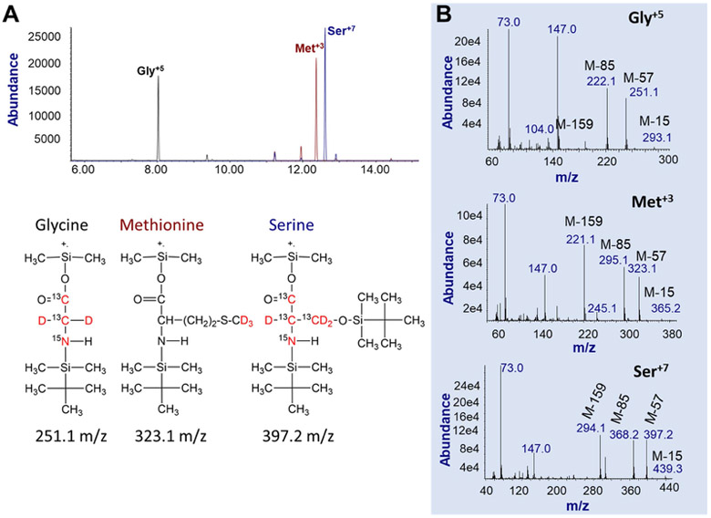
Figure 2. The GC/MS ion chromatograms and mass spectra of internal standard amino acids.
A. Selective ion monitoring (SIM) using the precursor (M+.)-57 ions. Show is the total-ion-current (TIC) elution profile of internal standard stable-isotope labeled (IS) amino acids Gly+5, Met+3, and Ser+7. The chemical structures of the three IS amino acids are presented below the TIC. Red colored atoms are the stable-isotope labeled atoms.
B. The full-scan spectra of IS amino acids including Gly+5 (top panel), Met+3 (middle panel) and Ser+7 (bottom panel). While molecular ions are not present, major fragmented ions are M+.-15, M+.-57, M+.-85, and M+.-159. In this study, M+.-57 ions were used for quantification using the SIM mode.

