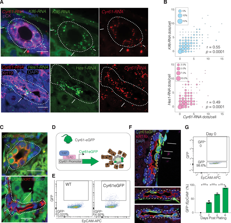
Figure 2. YAP Activity Defines BEC Heterogeneity In Vivo and Reflects a Dynamic Cell State In Vitro.
(A) Cyr61-Klf6 and Cyr61-Hes1 RNA-ISH combined with IF stain for pan-Cytokeratin (pCK) of mouse liver sections. Arrows indicate BECs co-expressing Cyr61 and Klf6 RNA, and arrowheads RNA-negative molecules.
(B) Bubble plots depicting the correlation of co-localized Cyr61-Klf6 and Cyr61-Hes1RNA molecules per BEC. Size of bubble corresponds to the respective co-expression frequency with inset showing size of bubble corresponding to % of cells with indicated frequency (n = 4 mice, BECs from 5 portal fields each, Spearman correlation).
(C) IF for pERK and JUNB (arrows) demonstrate heterogeneity within murine Cytokeratin19+ (KRT19+) BECs.
(D) Schematic for the Cyr61eGFP transgenic allele which expresses eGFP under the Cyr61 promoter and is used as a reporter for YAP transcriptional activity.
(E) Representative FACS analysis of GFP expression in BECs of a wildtype (WT) and a Cyr61eGFP mouse, where typically between 3–11% GFP+ BECs are seen.
(F) IF for GFP/KRT19 demonstrating clear intraductal heterogeneity of expression in the liver of a Cyr61eGFP-reporter mice. Arrows designate GFP+ cells.
(G) FACs analysis of freshly isolated GFP− BECs sorted from Cyr61eGFP mice, used for in vitro organoid growth assay. Bar plot depicts % of GFP+ cells over time, from 5000 initially seeded GFP− cells, showing that >90% of cultured BECs start expressing GFP within 6 days. (Mean ± SD, n = 3 mice, each in triplicate, ANOVA, followed by Tukey multiple comparisons test).
Dashed lines generally outline biliary structures.
See also Figure S2.

