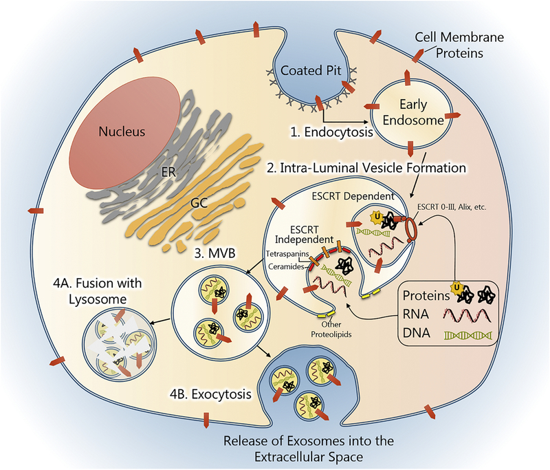
Figure 2. Exosome biogenesis.
Exosome biogenesis begins with endocytosis of the cell membrane via receptor-mediated endocytosis, clathrin-coated pits, pinocytosis, etc. This process creates a membrane-bound vacuole called an endosome. The endosomal membrane contains components of the cell membrane, such as receptors and signaling proteins, which were captured during endocytosis. Endosomes form intra-luminal vesicles (ILV), exosomes, when their membranes undergo subsequent invagination and scission. This process is facilitated by endosomal sorting complex required for transport (ESCRT) proteins or ESCRT-independent pathways, which are mediated by tetraspanins and proteolipids. During ILV formation, proteins and nucleic acids are selectively loaded into vesicles. Endosomes containing ILVs, also called multi-vesicular bodies (MVBs), can then merge with a lysosome and have their contents destroyed or be shuttled to the cell membrane and released.

