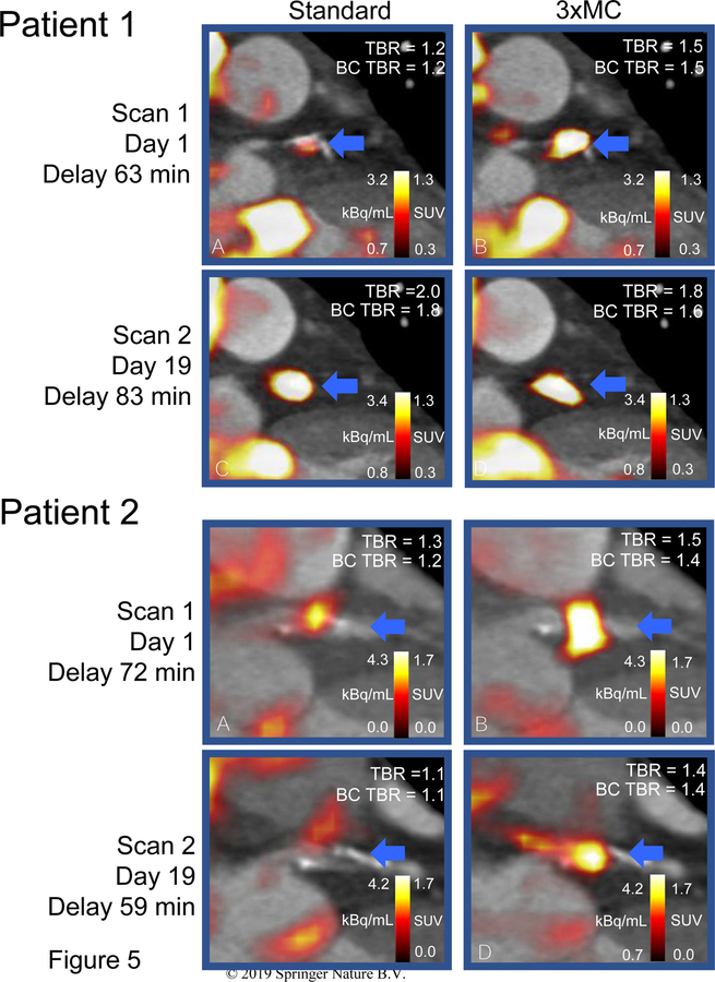
Figure 5: Test-rest coronary PET reproducibility before and after corrections.
Patient 1. Patient with significant respiratory and gross patient motion during the first scan (10.3 mm) and a 20-minute difference in the injection-to-scan delay leading to discrepant evaluations in the test-retest scans. Patient 2. Patient with several repositioning events (gross patient motion) during the acquisition, which in combination with the cardiorespiratory motion reduced the appearing tracer-uptake in the lesion. Both patients. In both cases 3×MC+BC reduced the intra-scan variability TBR evaluation of the lesion. Following 3×MC+BC the test-retest lesion evaluation was concordant (18F-NaF-avid) in comparison to discordant test-retest evaluations obtained from the standard images in both cases. TBR = target to background value, BC = blood pool correction. Standard = end-diastolic imaging, 3×MC = cardiorespiratory and gross patient motion corrected.

