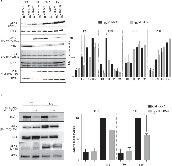
Figure 3.
Integrin α11 cytoplasmic tail contributes to FAK and ERK activation. (A) Serum-starved Huα11-WT and Huα11-1171 cells were plated on collagen I in serum-free conditions and cells were lysed at different time points (T0, T30, T45 and T60). Total and phosphorylated levels of FAKY397, ERK, p38, JNK were detected by Western blotting and the protein bands were quantified by densitometry analysis (full size immunoblots are shown in supplementary). (B) Human gingival fibroblasts (hGFs) were transfected with control (ctrl) siRNA or α11 siRNA (SMARTpool) and 48 hours post transfection, cells were serum-starved and plated on collagen I in serum-free conditions. After 30 mins, cells were lysed, and the lysates were analyzed by western blotting. Protein bands were quantified by densitometry analysis. Statistical significance was assessed by two tailed, unpaired t-tests and P-values are expressed as ***P < 0.001; **P < 0.01 and *P < 0.05.

