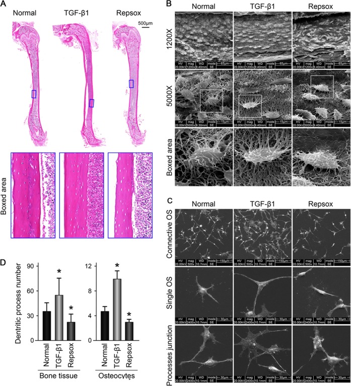
Fig. 1. TGF-β1 promotes the number of dendritic processes of osteocytes in bone tissue and MLO-Y4 cell line.
a Representative histological examination of cortical femora stained with HE showing lacuna size change in bone with TGF-β1 (10ng/ml) or its inhibitor Repsox (50 μM) after 21 days ex vivo culture (n = 6). b Representative SEM images revealing osteocyte canalicular system change in the cortex of mice treated with TGF-β1 (10 ng/ml) or Repsox (50 μM). Intact femurs were harvested from 4-week old mice and ex vivo cultured in 10% FBS DMEM with TGF-β1 or Repsox for 21 days (n = 6). Original magnification, ×1200 and ×5000. Details of the dendritic processes changes were shown in the boxed area (white). c Representative SEM images demonstrating the changes of dendritic processes of osteocytes (MLO-Y4 cell line) treated with TGF-β1 (5 ng/ml) or Repsox (25 μM). The cells were incubated with TGF-β1 or Repsox for 24 h. Accordingly, process junctions between cytoplasmic arms of osteocytes altered (lower lane). The results shown are based on three independent experiments (n = 3). d Quantitative analyses of the dendritic processes changes of osteocytes in b, c. Quantitative analyses were based on three independent experiments (n = 3). The results are shown as the mean ± s.d.; n = 3; *p < 0.05 by t-test

