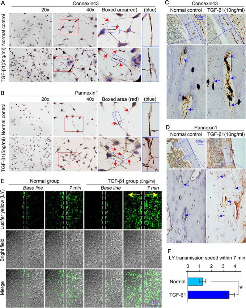
Fig. 3. TGF-β1 promotes the connectivity between osteocytes via increasing the expression of Cx43 and pannexin1.
a, b Representative images of immunohistochemistry (IHC) using the anti-Cx43 antibody (top) and anti-pannexin1 antibody (bottom) coupled with Gill’s haematoxylin counterstaining on cells cultured on glass slides showing increased expression of Cx43 and pannexin1 after TGF-β1 (5 ng/ml) treatment. Boxed area (red) shows that Cx43 and panx1 clustered to form plaques at the sites which eventually lead to gap junction formation. Additionally, panx1 also showed its expression on the membrane and in the cytoplasm at the tips of dendritic processes. The results shown are based on three independent experiments (n = 3). c, d Representative immunohistochemistry staining (IHC) images confirming the enhanced expression of Cx43 (up) and panx1 (down) in osteocytes residing in cortical bone of femur after treatment with TGF-β1(10 ng/ml). The results shown are based on three independent experiments (n = 3). e The Lucifer yellow (LY) coupling assay showing the increased transmission of dye (small fluorescent molecules) between living osteocytes (MLO-Y4 cell line) induced by TGF-β1 (5 ng/ml) by CLSM at ×40 magnification. The dye transmission images were obtained at 7 min after Lucifer yellow stain in the living osteocytes. The results shown are based on three independent experiments (n = 3). f Statistical analysis showing the different transmission speeds of Lucifer yellow between the control and TGF-β1 (5 ng/ml) group. Data are presented as the mean ± s.d.; n = 3, *p < 0.05 by t-test

